Neuroimmunology · Meningeal immunity

How do researchers study meningeal immunity and neuroimmune interfaces in preclinical models?

Studying meningeal immunity, dural lymphatics, and immune surveillance at the brain border requires imaging tools that match the biological question — from live cell dynamics to whole-cranium anatomy. Neuroimmunology labs today draw on a mix of ex vivo and in vivo methods, each with distinct strengths.

What researchers typically do

Much of modern meningeal immunity research builds on ex vivo approaches: dural whole mounts with fluorescent staining imaged by light or confocal microscopy, histology and immunohistochemistry of meningeal tissue, and — for live dynamics — intravital two-photon microscopy through thinned skull windows. Dural whole mounts necessarily separate the dura from the brain and skull, so spatial relationships at the brain border are lost. Intravital two-photon preserves live cell behavior but is typically limited to the thinned region and a finite imaging depth. These are established, valuable tools — and many labs continue to use them alongside newer approaches. Histology, whole-slide imaging, and light-sheet microscopy are also common in neuroimmunology studies, usually analyzed separately from any cranial reference volume.

In situ cranial mapping for neuroimmune discovery

The iterative in situ micro-computed tomography (micro-CT) workflow published in Cell Reports Methods (Rosenblum et al., 2021) and STAR Protocols (Dang et al., 2023) is the only published method to map skull, brain, and meningeal vasculature together in the same intact head — preserving the anatomical context that dural whole mounts and localized intravital windows cannot. The NeuroSimplicity Anatomic Imaging Module works seamlessly with this workflow — and is the only analysis tool available that does. NIH NINDS collaborators used this pipeline in Nature (2024, 2026) to map vascular connectomics and meningeal lymphatics at the brain border. When labs license the full Imaging Suite (Anatomic Imaging, Molecular Imaging, Digital Pathology, and Spatial Omics Modules together), they can further register histology, whole-slide imaging, light-sheet, and other modalities from the same cohort into one native sample-space framework.

What the platform enables

  • In situ cranial mapping of meningeal lymphatics, dural sinuses, and venolymphatic structures
  • Iterative in situ micro-computed tomography (micro-CT) via the Anatomic Imaging Module
  • Cross-modal registration across the full Imaging Suite — histology, whole-slide imaging, light-sheet, and confocal from the same cohort
  • Atlas registration to the sample (e.g., Allen Atlas CCFv3, NeuroSimplicity Cranial and Vessel Atlases)
  • Automated feature extraction, labeling, and quantitative metrics in native sample space

More detail

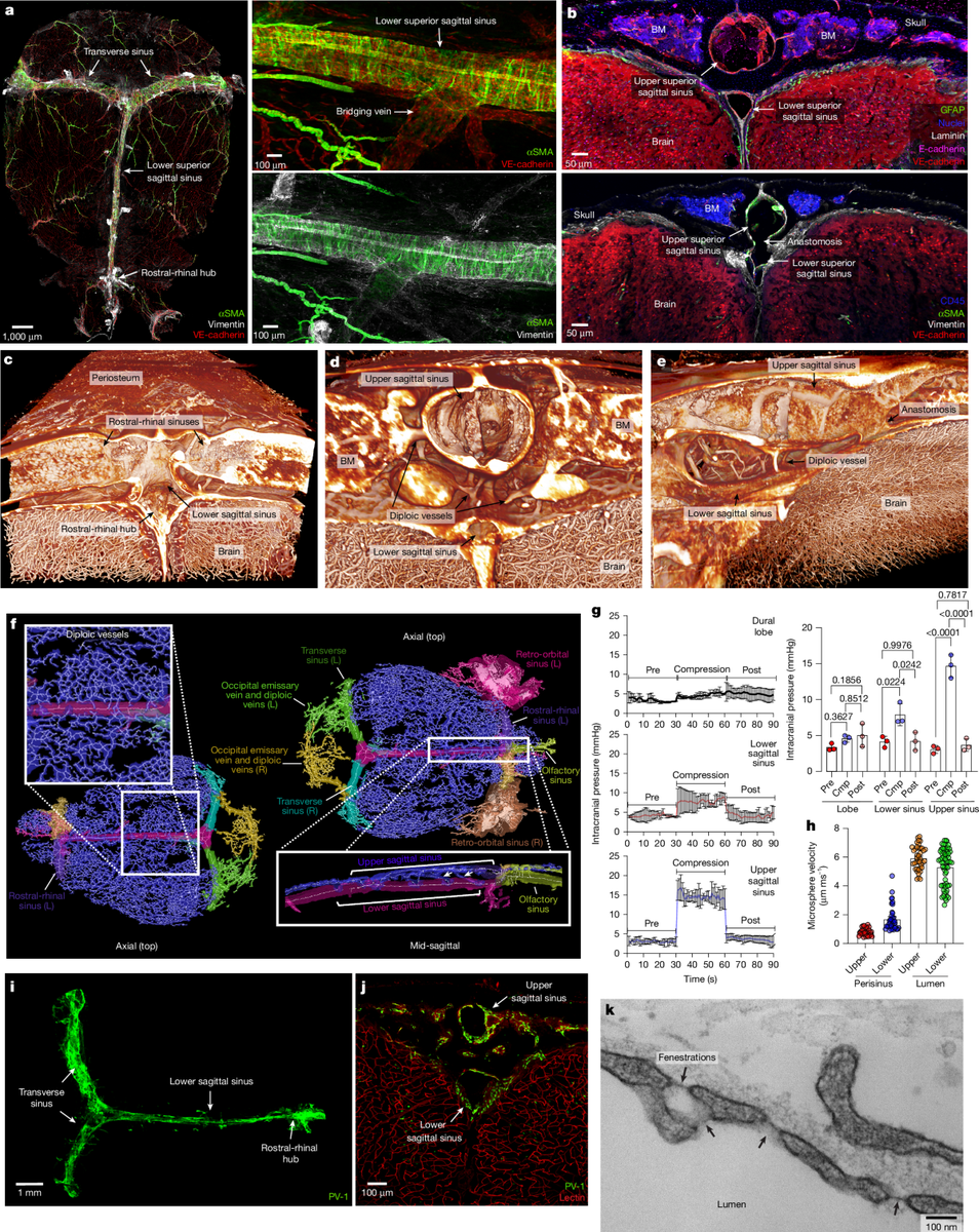
Cranial vasculature mapping from Nature (2026) — dural sinuses and meningeal immunity at the brain border

NIH NINDS collaborators applied the in situ neurovascular workflow in Nature (2024, 2026) to map vascular connectomics, meningeal lymphatics, dural sinuses, venolymphatic hubs, and dural-associated lymphoid tissues (DALT) at the brain border.

The underlying in situ micro-CT workflow is documented in Cell Reports Methods and STAR Protocols. For bench adoption, see the neurovascular workflow guide and protocol links in Next steps below.

Peer-reviewed research

This workflow is documented in Cell Reports Methods and STAR Protocols, and applied in Nature discovery studies by NIH NINDS collaborators. Full citations, figures, and paper links are on our publications page.

View all publications

Next steps

Neuroimmunology Imaging Tools — Meningeal Immunity & Brain Border Studies | NeuroSimplicity