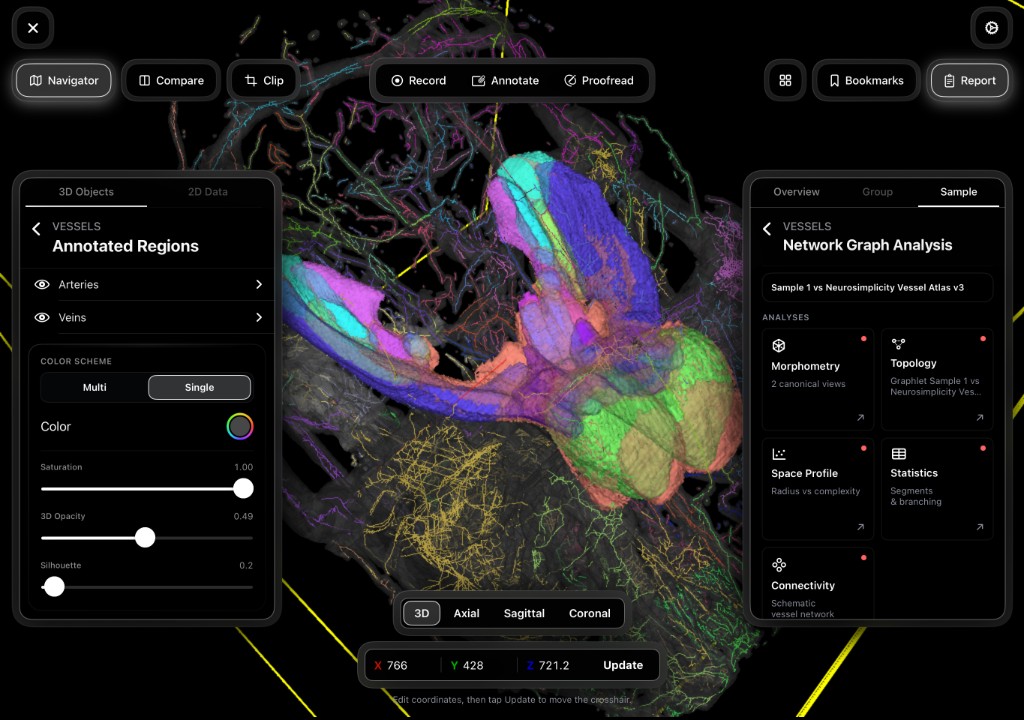
How can I map the brain, skull, and vessels without destroying my sample?
Combine multi-modal data to explore neurovascular interfaces in situ — intact and quantifiable.
Explore this workflowMultimodal preclinical neuroscience data processing platform — regional quantification and multi-modal integration on your on-premises private cloud.
Used in landmark studies published in Nature and Cell journals by NIH researchers.
Institutional Control | Enterprise Performance
The Imaging Suite is the core platform — buy the modules you need, connect labs, visualize and quantify together.
Reliably integrate neuroscientific data across all scales—from anatomic to molecular.
Deploy on your infrastructure with hybrid on-premises private cloud — without sending data off-site.
Toggle modalities in the sample-space visualizer below.
Toggle modalities, fly through sample-space reconstructions, and explore regional quantification outputs.
Loading viewer
Navigator
3D Objects
View presets
Built for giga-scale preclinical datasets your team already generates. Compare cohorts, export publication-ready views, and share securely across labs on your own infrastructure.
Lab sharing
Private cloud
Air-gapped ready
Outputs include vessel diameter distributions, regional cell counts, connectivity graphs, and atlas-labeled metrics — all in native sample space.
Micro-CT, light sheet, pathology, and spatial omics — plan the modules your lab needs. Each integrates in native sample space on your on-premises private cloud.
Peer-reviewed research demonstrating the Imaging Suite's capabilities in advancing neuroscientific discovery.
Combine multi-modal data to explore neurovascular interfaces in situ — intact and quantifiable.
Explore this workflow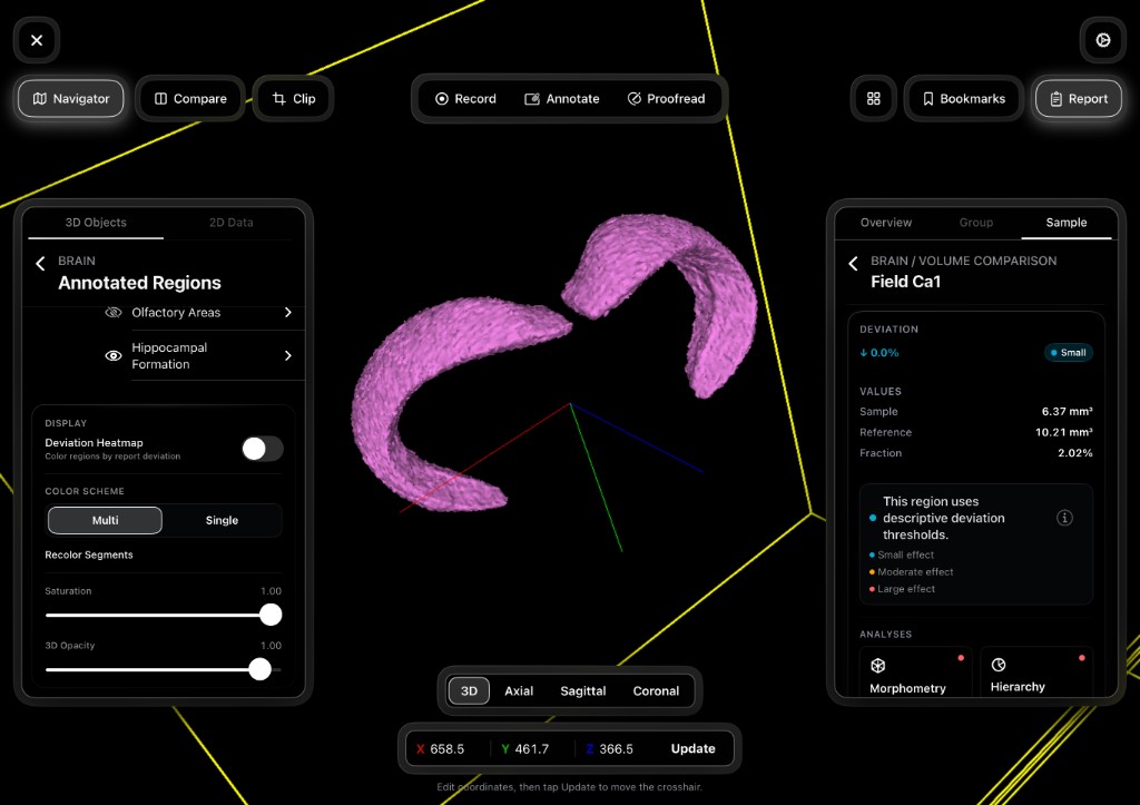
Register atlases to the sample, not the other way around, so regional metrics reflect true geometry.
Explore this workflow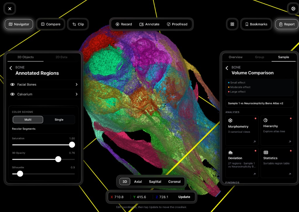
Explore automatically generated 3D regional morphometrics while keeping the intact anatomic context.
Explore this workflow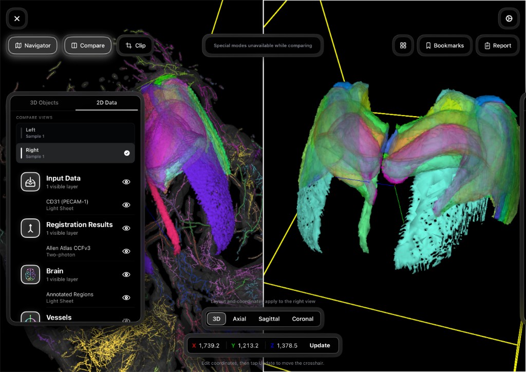
Compare samples and atlases side-by-side, exploring automatically generated quantitative metrics and 3D visualizations.
Explore this workflow| Benchmark | The Imaging Suite | Legacy Standard |
|---|---|---|
| Reproducibility | Deterministic | Stochastic |
| Data Control | Institutional Control | Third-Party Dependent |
| Cohort Comparisons | Automated | Manual Intervention |
| Operational Cost | High-Efficiency OpEx | Linear Scale Pricing |